Abiocode Logo
Products
Contact Us
- Telephone:
1-818-707-0309 - E-Mail:
Abiocode@Abiocode.com
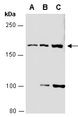
R1423-2 - MAP3K4 (C) Antibody, Rabbit Polyclonal
|
Quantity: 100 ul Application: WB
Predicted I Observed M.W.: 182 kDa Uniprot ID: Q9Y6R4 Background: The central core of each mitogen-activated protein kinase (MAPK) pathway is a conserved cascade of 3 protein kinases: an activated MAPK kinase kinase (MAPKKK) phosphorylates and activates a specific MAPK kinase (MAPKK), which then activates a specific MAPK. While the ERK MAPKs are activated by mitogenic stimulation, the CSBP2 and JNK MAPKs are activated by environmental stresses such as osmotic shock, UV irradiation, wound stress, and inflammatory factors. MAP3K4 contains a protein kinase catalytic domain at the C terminus. The N-terminal nonkinase domain may contain a regulatory domain. Expression of MAP3K4 in mammalian cells activated the CSBP2 and JNK MAPK pathways, but not the ERK pathway. In vitro kinase studies indicated that recombinant MAP3K4 can specifically phosphorylate and activate PRKMK6 and SERK1, MAPKKs that activate CSBP2 and JNK, respectively but cannot phosphorylate PRKMK1, an MAPKK that activates ERKs. MAP3K4 is a major mediator of environmental stresses that activate the CSBP2 MAPK pathway, and a minor mediator of the JNK pathway. Other Names: Mitogen-activated protein kinase kinase kinase 4, MAP three kinase 1, MAPK/ERK kinase kinase 4, MEK kinase 4, MEKK 4, KIAA0213, MAPKKK4, MEKK4, MTK1 Source and Purity: Rabbit polyclonal antibodies were produced by immunizing animals with a GST-fusion protein containing the C-terminal region of human MAP3K4. Antibodies were purified by affinity purification using immunogen. Storage Buffer and Condition: Supplied in 1 x PBS (pH 7.4), 100 ug/ml BSA, 40% Glycerol, 0.01% NaN3. Store at -20 °C. Stable for 6 months from date of receipt. Tested Applications: WB: 1:1,000-1:5,000 (detect endogenous protein*) *: The apparent protein size on WB may be different from the calculated M.W. due to modifications. Species Specificity: Human, Mouse Product Data:
|