Abiocode Logo
Products
Contact Us
- Telephone:
1-818-707-0309 - E-Mail:
Abiocode@Abiocode.com
R3421-1 - PIK3CD (N) Antibody, Rabbit Polyclonal
|
Quantity: 100 ul Application: WB
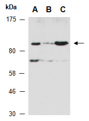
Predicted I Observed M.W.: 119 kDa Uniprot ID: O00329 Background: Phosphatidylinositol 4,5-bisphosphate 3-kinase catalytic subunit delta isoform (PIk3CD) is a phosphoinositide-3-kinase (PI3K) that phosphorylates PftdIns(4,5)P2 (Phosphatidylinositol 4,5-bisphosphate) to generate phosphatidylinositol 3,4,5-trisphosphate (PIP3). PIP3 plays a key role by recruiting PH domain-containing proteins to the membrane, including AKT1 and PDPK1, to activate signaling cascades involved in cell growth, survival, proliferation, motility and morphology. PIK3CD mediates immune responses. PIK3CD is required for B-cell receptor (BCR) signaling and plays a role in B-cell development, proliferation, migration, and function. PIK3CD is also required for T-cell receptor (TCR) signaling and proliferation, signaling and cytokine production of naive, effector and memory T-cells. Together with PIK3CG, PIK3CD is involved in natural killer (NK) cell development, maturation and function. Other Names: Phosphatidylinositol 4,5-bisphosphate 3-kinase catalytic subunit delta isoform, PI3-kinase subunit delta, PI3K-delta, PI3Kdelta, PtdIns-3-kinase subunit delta, Phosphatidylinositol 4,5-bisphosphate 3-kinase 110 kDa catalytic subunit delta, PtdIns-3-kinase subunit p110-delta, p110delta Source and Purity: Rabbit polyclonal antibodies were produced by immunizing animals with a GST-fusion protein containing the N-terminal region of human PIK3CD. Antibodies were purified by affinity purification using immunogen. Storage Buffer and Condition: Supplied in 1 x PBS (pH 7.4), 100 ug/ml BSA, 40% Glycerol, 0.01% NaN3. Store at -20 °C. Stable for 6 months from date of receipt. Tested Applications: WB: 1:500-1:2,000 (detect endogenous protein*) *: The apparent protein size on WB may be different from the calculated M.W. due to modifications. Species Specificity: Human, Mouse Product Data: |